“It can scan blood vessels similar to the thickness of a human hair” Development of contrast medium looking through blood vessels 10 times minutely
The contrast medium developed has an effect for long-term MRI scan
![The pictures of brain blood vessels taken by using the existing contrast medium and new contrast medium [Photo provided = The Institute for Basic Science]](/news_img/73/257c86617c9540faff211203a64d6494.jpg)
The pictures of brain blood vessels taken by using the existing contrast medium and new contrast medium [Photo provided = The Institute for Basic Science]
The contrast medium that can look through blood vessels 10 times minutely than the existing contrast medium has been developed and early diagnosis of vascular diseases is available.
The Institute for Basic Science (IBS) Center for Nanomedicine held the development of new type of contrast medium performance briefing on the 8th afternoon in Seodaemun-gu Seoul and announced a joint research team of prof. Jin-woo Cheon Department of Chemistry at Yonsei University (Director of IBS Center for Nanomedicine) and prof. Byeong-wook Choi Yonsei University College of Medicine developed contrast medium that can look through blood vessels 10 times minutely than the existing contrast medium.
The new contrast medium named SAIO (Supramolecular Amorphous-like Iron Oxide) by the research team is about 5㎚ size (Nanometer one billionth (short scale) of a meter) in the form of carbohydrate (Dextran) nanoparticles coated with iron. The particle is 1500 times smaller than micro vessels so it can get around the vessels.
Director Cheon said “If the current MRI technology is in the level where it sees only big highways MRI scan taken by using SAIO can observe narrow side streets in detail. Through this we have succeeded in developing a precise 3-dimension blood vessels map.”
According to this briefing the existing contrast medium (gadolinium-based) shows 1㎜ of resolution but the new iron-based contrast medium can scan 10 times smaller particles.
The reason why the new contrast medium can make clearer scans than the typical one is because it has longer residence time. Gadolinium-based contrast medium has short residence time so long-term MRI scan was difficult. However the new contrast medium shows relatively long angiography effect. MRI has been known that the more time it spends on scanning the more precise scans can be gained.
The researchers took MRI scans of animals’ brains (Rats) to see the effect of angiography. As a result it was able to scan micro vessels with the thickness of a human hair (100㎛).
Furthermore this SAIO contrast medium was confirmed during the animal test that it is completely emitted in the urine. Director Cheon explained “The new contrast medium can pass through the filter in kidney glomerulus” and added “In 8 hours 92% is excreted after the scanning and in 24 hours most of them is excreted.”
Prof. Byeong-wook Choi said “It is expected to increase the diagnostic accuracy of brain cardiovascular diseases such as stroke myocardial infarction angina and dementia.”
This research was conducted with the supports from the IBS the leading research-centered hospitals project from the Ministry of Health and Welfare and the Ministry of Science and ICT and Dr. Tae-hyeon Shin Dr. Pan-ki Kim and researcher Seong-hwi Kang participated. The thesis was published in ‘Nature Biomedical Engineering’ (IF 18.952) on March 9th.
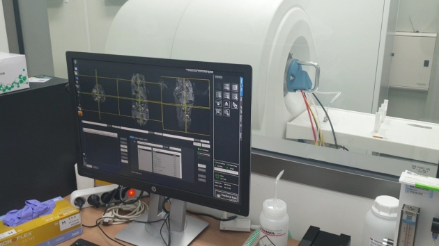
The result of scanning rat with MRI equipment and MRI equipment for animals
![The contrast medium that can look through blood vessels 10 times minutely than the existing contrast medium has been developed by the IBS Center for Nanomedicine. (From the left) researcher Seong-hwi Kang Dr. Tae-hyeon Shin prof. Jin-woo Cheon prof. Byeong-ok Choi Dr. Pan-ki Kim and researcher Jeong-soo Na [Photo provided = The Institute for Basic Science]](/news_img/73/4411a4a1534dc850bdd615372a4b5e77.jpg)
The contrast medium that can look through blood vessels 10 times minutely than the existing contrast medium has been developed by the IBS Center for Nanomedicine. (From the left) researcher Seong-hwi Kang Dr. Tae-hyeon Shin prof. Jin-woo Cheon prof. Byeong-ok Choi Dr. Pan-ki Kim and researcher Jeong-soo Na [Photo provided = The Institute for Basic Science]
Website: : http://www.wip-news.com/news/articleView.html?idxno=5558